Diet and Nutrition

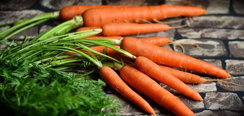
Adding powerful antioxidants to your diet can improve your eye health.

Did you know that small dietary changes can have a big impact on your eyes? Adding certain nutrients to your daily diet—either through foods or supplements—can help preserve your vision. Researchers have linked eye-friendly nutrients, such as lutein and zeaxanthin, vitamin C, vitamin E and zinc, to reducing the risk of certain eye diseases.

Lutein and Zeaxanthin
Lutein and zeaxanthin are important nutrients found in green leafy vegetables such as spinach or kale, as well as other foods, such as eggs. Many studies show that lutein and zeaxanthin reduce the risk of chronic eye diseases, including age-related macular degeneration and cataracts.

Vitamin C
Vitamin C (ascorbic acid) is an antioxidant found in fruits and vegetables. Scientific evidence suggests vitamin C lowers the risk of developing cataracts. Also, when taken in combination with other essential nutrients, it can slow the progression of age-related macular degeneration and visual acuity loss.

Vitamin E
Vitamin E is a powerful antioxidant found in nuts, fortified cereals and sweet potatoes. Research indicates it protects cells in the eyes from unstable molecules called free radicals, which break down healthy tissue.

Essential Fatty Acids
Fats are a necessary part of the human diet. They maintain the integrity of the nervous system, fuel cells and boost the immune system. Research shows omega-3 fatty acids are important for proper visual development and retinal function.

Zinc
Zinc is an essential trace mineral or “helper molecule.” It plays a vital role in bringing vitamin A from the liver to the retina in order to produce melanin, a protective pigment in the eyes. Zinc is highly concentrated in the eye, mostly in the retina and choroid, the vascular tissue layer lying under the retina.

Age-Related Macular Degeneration (AMD)

Age-Related Macular Degeneration is the leading cause of severe vision loss in people over age 50. The Centers for Disease Control and Prevention estimate that 1.8 million people have AMD and another 7.3 million are at substantial risk for vision loss from AMD.

There may not be any signs or symptoms in the early stages of macular degeneration which is why yearly examinations by your eye care professional is recommended. Symptoms can include gradual loss of the ability to see objects clearly or appear distorted in shape. Straight lines look wavy or appear crooked. Loss of clear color vision or a dark or empty area appears in the center of vision.

This eye disease occurs when there are changes to the macula, a small portion of the retinal that is located on the inside back layer of the eye. AMD is a loss of central vision that can occur in tow forms: “dry” or atrophic and “wet” or exudative.

There is no cure for dry AMD and any loss in central vision cannot be restored. Recent research shows a possible link between nutrition and the progression of dry AMD. Dietary changes favoring low-fat content and dark green leafy vegetables can slow vision loss and nutritional supplements may be beneficial.

Although less common “wet” macular degeneration results when fluids leak from newly formed blood vessels under the macula and blur central vision. Vision loss can be rapid and severe. Early detection is critical and can be treated with laser treatments or other medications that are injected directly into the back of the eye. These are not permanent cures but are used to slow the rate of central vision loss.

Children’s Vision

Childrens Vision

Vision doesn’t just happen. A child’s brain learns how to use eyes to see just like it learns how to use legs to walk or a mouth to learn how to speak. Children with uncorrected vision conditions face many barriers in life. The longer a vision problem goes undiagnosed and untreated the more difficult it becomes to overcome these barriers.

As children progress through school they face increasing demands on their visual abilities. Every child needs to have the vision skills for effective reading and learning. Vision is more than visual acuity, the ability to see clearly and eye focusing, ability to quickly and accurately maintain clear vision as the distance from objects change. Other visual perceptual skills include recognition, comprehension, retention, eye tracking, eye teaming and hand eye coordination.

A child may not tell you that he or she has a vision problem because they may think the way they see in normal. Signs that may indicate a vision problem may include frequent eye rubbing or blinking, avoiding reading and other near activities, frequent headaches, covering one eye, seeing double, holding materials close to the face and tilting the head to one side.

Some children with learning difficulties exhibit specific behaviors of hyperactivity and distractibility and are often labeled as having Attention Deficit Hyperactivity Disorder (ADHD). If a child’s visual skills are lacking they can experience short attention span, letters or words jumble together, loss of place when reading and difficulty remembering what he or she read.

Vision changes can occur without your child or you noticing them. Therefore, your child should receive an eye examination every year as recommended by your eye doctor. Early detection and treatment provide the very best opportunity to correct vision problems to make sure your child has the best possible tools to learn successfully.

Cataracts

Cataracts

The simplest definition of a cataract is that it is a clouding of the lens of your eye. The lens is normally clear in a healthy eye. This clouding normally develops slowly over time and symptoms may at first be nearly imperceptible. Cloudiness may start out in only a small part of the eye and may not initially be noticeable. They will, however, usually become more advanced over a period of time.

Cataracts may cause a change of color in one or both of the eyes. Your eyes may begin to look yellowish or even light brown in color. Eyes that are sensitive to light are sometimes one of the first signs that you have cataracts. Lamps, car headlights, and even sunlight that didn’t seem to bother you before, may suddenly seem to be too bright. If you are experiencing increased problems seeing well and driving safely when it is dark, it is probably a good idea to get tested for cataracts.

If you find that you need to increase your contact or glasses strength on a frequent basis this could be an indication that you have cataracts. Seeing double images could be a sign of several serious health problems as well as a symptom of cataracts. Monocular double vision can cause several images to appear in one of the eyes, but not the other eye. Multiple images that occur in one eye are often caused by cataracts.

Difficulty seeing colors is another symptom of having cataracts. Colors may appear to fade and not be as vibrant as they once were. There can also be a yellowing of colors that are seen. When suffering from cataracts it may seem as if there is a glaze or a permanent film that is covering the eyes. It might feel as if a sheer, transparent layer is covering one or both of the eyes.

Glaucoma

Glaucoma is a group of eye disorders that lead to progressive damage to the optic nerve. People with glaucoma can lose nerve tissue, resulting in vision loss. The optic nerve is a bundle of about 1 million individual nerve fibers that transmits the visual signals from the eye to the brain. In the most common form of glaucoma, primary open-angle glaucoma, the fluid pressure inside the eye increases. This increase in pressure may cause progressive damage to the optic nerve and loss of nerve fibers. Vision loss may result. Advanced glaucoma may even cause blindness.

Glaucoma is the second-leading cause of blindness in the U.S. It most often occurs in people over age 40,although an infant (congenital) form of glaucoma exists. People with a family history of glaucoma, African Americans over the age of 40 and Hispanics over the age of 60 have an increased risk of developing glaucoma. Other risk factors include thinner corneas, chronic eye inflammation and taking medications that increase the pressure in the eyes.

The most common form of glaucoma, primary open-angle glaucoma, develops slowly and usually without any symptoms. Many people are not aware they have the condition until they have significant vision loss. Initially, glaucoma affects peripheral or side vision, but it can advance to central vision loss. If left untreated, glaucoma can lead to significant vision loss in both eyes, and may even lead to blindness.

A less common type of glaucoma, acute angle-closure glaucoma, usually occurs abruptly due to a rapid increase of pressure in the eye. Its symptoms may include severe eye pain, nausea, redness in the eye, seeing halos or colored rings around lights and blurred vision. This is an emergency condition in which severe vision loss can occur quickly; see your optometrist immediately.

Glaucoma cannot currently be prevented. But if it is diagnosed and treated early, it can usually be controlled. Medication or surgery can slow or prevent further vision loss. However, vision already lost to glaucoma cannot be restored. That is why the American Optometric Association recommends an annual dilated eye examination for people at risk for glaucoma. Depending on your specific condition, your doctor may recommend more frequent examinations.

Conjunctivitis

Conjunctivitis

Conjunctivitis is an inflammation or swelling of the conjunctiva. The conjunctiva is the thin transparent layer of tissue that lines the inner surface of the eyelid and covers the white part of the eye.

Often called “pink eye,” conjunctivitis is a common eye disease, especially in children. It may affect one or both eyes. Some forms of conjunctivitis are highly contagious and can easily spread in schools and at home. While conjunctivitis is usually a minor eye infection, sometimes it can develop into a more serious problem.

A viral or bacterial infection can cause conjunctivitis. It can also develop due to an allergic reaction to air irritants such as pollen and smoke, chlorine in swimming pools, ingredients in cosmetics, or other products that contact the eyes, such as contact lenses. Sexually transmitted diseases like chlamydia and gonorrhea are less common causes of conjunctivitis.

People with conjunctivitis may experience symptoms of a gritty feeling, itching, burning, excessive tearing, swollen lids, discharge from one or both eyes, red eyes and increased sensitivity to light.

Conjunctivitis can be diagnosed through a comprehensive eye examination with special emphasis on the conjunctiva and surrounding tissues. The appropriate treatment for conjunctivitis depends on its cause. Using the information obtained from these tests, your optometrist can determine if you have conjunctivitis and advise you on treatment options.

Diabetic Retinopathy

Diabetic Retinopathy
According to the American Diabetes Association nearly 21 million Americans are affected by diabetes and another 54 million are considered pre-diabetic. It is the leading cause of death, disability and blindness in the US for adults ages 20-74. The CDC estimates that 4.1 million people currently have diabetic retinopathy and that by the year 2020 7.2 million will have the eye disease.

Diabetic retinopathy causes progressive damage to the retina, the light-sensitive lining at the back of the eye. Diabetic retinopathy is a serious sight-threatening complication of diabetes. Diabetes interferes with the body’s ability to use and store sugar (glucose). The disease is characterized by too much sugar in the blood, which can cause damage throughout the body, including the eyes.

Over time, diabetes damages the blood vessels in the retina. Diabetic retinopathy occurs when these tiny blood vessels leak blood and other fluids. This causes the retinal tissue to swell, resulting in cloudy or blurred vision. The condition usually affects both eyes. The longer a person has diabetes, the more likely they will develop diabetic retinopathy. If left untreated, diabetic retinopathy can cause blindness.

Symptoms of diabetic retinopathy include seeing spots or floaters, blurred vision, having a dark or empty spot in the center of your vision, difficulty seeing at night and vision that seems to fluctuate. When people with diabetes experience long periods of high blood sugar, fluid can accumulate in the lens inside the eye that controls focusing. This changes the curvature of the lens, leading to blurred vision and or changes in a glasses prescription. This is commonly referred to as a diabetic shift.

Treatment for diabetic retinopathy depends on the stage of the disease. The goal of any treatment is to slow or stop the progression of the disease. In the early stages of non-proliferative diabetic retinopathy, regular monitoring may be the only treatment. If the disease advances, the blood vessels can leak blood and fluid into the retina, leading to macular edema. Laser treatment and medication injected directly into the back of the eye can stop or slow down the progression of this leakage.

If you are diabetic, you can help prevent or slow the development of diabetic retinopathy by taking your prescribed medication, maintaining a proper diet, exercising regularly, controlling high blood pressure and avoiding alcohol and smoking. Following your doctor’s advice for controlling sugar levels can help control the progression of the disease and reduce the risk of diabetic retinopathy by as much as 76 percent. It is recommended to have annual dilated eye examinations to monitor the progress of this eye disease.